Phytopathology pictures (fungi, bacteria, viruses and so on)

Hello everyone,

In my previous post (Play raw: Sharpen_techniques_lizard_subject) Sebastien Guyader asked me some quesitons about my pictures and I prefer to open a new topic about this topic.

@sguyader
What is the white stuff in the right petri dish? Paper?

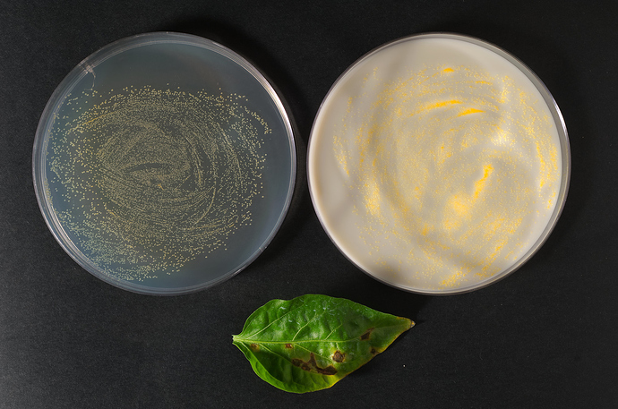
Here is my jpeg picture concerning a picture of a plant disease on pepper leaves caused by a bacterium which is Xanthomonas vesicatoria quite common in Northern Italy:

Here is the NEF raw file (Nikon D700):

This picture is difficult to improve because the bacterium is growing on a petri dish where there is also, as a component, Calcium Carbonate which is responsable of the white colour of the petri dish on the right (the dish on the left is different: CSGA medium).
Since the colour of the bacterium is yellow it is quite difficult for me to improve the picture in order to make this bacterium more visible in the white dish on the right side.
With RawTherapee I suppose I should try the Local editing branch in order to select only the white dish and try to work exclusively on it.
With Darktable I suppose I should opt for the mask route…
Needless to say, I could export this Raw image and work with Gimp as well :slight_smile:

2 Likes

Since Sebastian is both a mycologist and a virologist let me add 2 more pictures :slight_smile:

Since the previous disease concerns pepper let me continue on this plant :slight_smile:

Here is a picture regarding a virus, CMV (Cucumber mosaic virus) on pepper fruits.
As usual, I take my pictures with a Nikon D700, ISO 200, 2850 K (as manual white balance) with a 60 mm Nikkor micro lens 2,8 F. As lateral source of light, I work with a single lamp (Elinchrom system).

Here is the jpeg picture:

Here is the Raw file (Nikon D700) on Dropbox:

With this image, personally, I would decrease only the shadow on the left side of the pepper fruit.

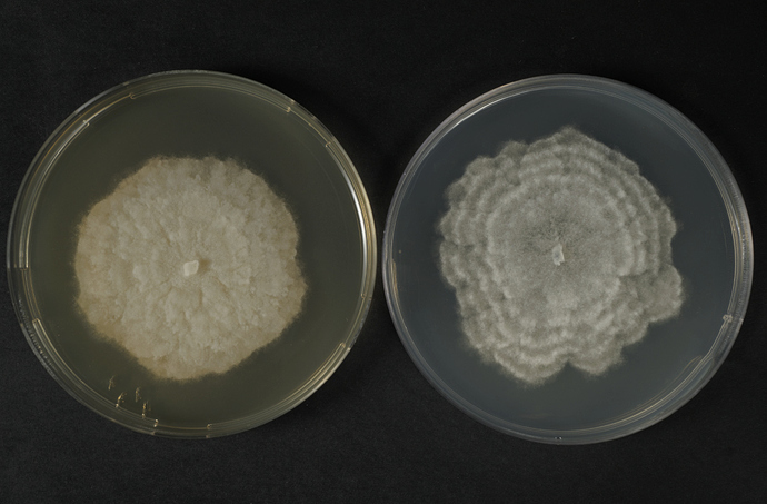
I add one more image of mine: a disease on pepper caused by a fungus which is Phytophthora capsici.
The picture displays the same fungus growing on 2 petri dishes at 10 days of growth. The first petri dish is MA (malt agar), the second one, on the right, PDA (potato dextrose agar).

Here is the jpeg image (quite compressed to post on this forum):

Here is the same image on Dropbox (Raw file - Nikon D700, 9 mb):

Personally, as regards this image, I would prefer to leave as it is right now :slight_smile:
Maybe just a bit of sharpening…

1 Like

Thanks @Silvio_Grosso.
Here are a couple of images of the fungus I’ve been studying these pas years:

Necrotic symptoms caused by the fungus Colletotrichum alatae on “white yam” (Dioscorea alata) leaf in the field:

And here a (bad) image of a microscopic view of a germinated spore on glass slide:

For the curious, the darker “bulbs” are called appressoria, they are a cellular structure which exert significant physical pressure (some have measured a pressure as high as 80 bars) as well as enzymatic digestion of leaf cuticle, which allow the germ tubes to forcibly penetrate inside the leaf tissues.

2 Likes

Helo @sguyader

I have worked extensively with Colletotrichum species, mostly related to apple fruits, even though I am far from being an expert on the subject…
BTW, nice pictures indeed :slight_smile:

1 Like

There is also the Color Toning > Color correction regions tool in RT 5.6. You can do things like this:


XANTHOMONAS_VESICATORIA_peperone_foglie__370-SFR_GYCA_CSGA_48H.jpg.out.pp3 (11.9 KB)

2 Likes

Hello @Thanatomanic

Thanks a lot indeed for your help since I didn’t know this tool of RawTherapee.
Your image is better than mine :slight_smile:

Just for nitpicking though, your result on my display makes the light yellow of these bacteria become an orange where the bacteria are more concentrated on the petri dish.
A more “orange colour” is more related to other bacteria (e.g. Erwinia herbicola).

Here is a different picture, always Xanthomonas on GYCA petri dish, where these bacteria show their “true” light yellow colour (at least on my display of course…):

@Silvio_Grosso I had no intention of accurately showing the colour of the colonies :slight_smile:, just to show the possibility of the tool.

I am curious though, what is the purpose of your photographs in relation to your research? You seem to have put some thought in your camera settings and lighting. Do you need to inspect the plants for the progression of their disease? Is color accuracy important? If so, have you photographed a color target (e.g. X-Rite ColorChecker)? And why use a single lamp?

Hello @Thanatomanic

Do you need to inspect the plants for the progression of their disease? Is color accuracy important? If so, have you photographed a color target

Our Italian public laboratory receives every year hundreds of samples with some unknown plant disease, mostly fungi, bacteria, viruses, nematodes and we are supposed to discover what is the “killer” (culprit) of these plants.
As a result, I am charged to keep track, as a photographer, of these samples even though my real day job concerns plant pathology, not photography :slight_smile:

In the past, I did have tried a X-Rite color checker but in the end, at least on my display of course, I have realized it was not completely necessary because the colours were pretty much accurate out of the box when I displayed the Raw files on my display with RawTherapee (Amaze method together with the standards settings) or other “similar” softwares (Windows 10 as system).

With a thermo colorimeter tool a colleague of mine measured the Kelvin light (temperature) of our Elinchrom lamp and he suggested me to always set accordingly my Nikon D700 with 2850 kelvin (manual white balance) which I duly do :slight_smile:

I keep the ISO as low as possible (usually 200) and I never take my picture with a very closed diafram (usually F8 - F9, as the uppermost closure).
The Nikon D700 is always mounted on a tripod and it is over my samples.

I have only always used a lamp so far (which is on my right) because, on the left (opposite to it), I have got a white sheet of paper to reduce the shadows of the samples (which lies in the middle of this setup). Quite often I replace the white sheet with a plastic panel (30x50 cm) with a “Silver” coat on it to decrease even further the shadows generated by my samples with the supplementar advantage of illuminating the sample on its left side.
BTW, in front of the lamp there is a huge “semi-transparent” sheet of paper to reduce the strenght of the light generated by this lamp.

Sorry but my technical English is not good enough to describe these tools exactly…

And yep, colour accuracy is KEY for my pictures (together with sharpness) :slight_smile:

EDIT: if you take a closer look at the white dish (the one on the right of the first picture) you can easily spot the shadows generated both by the other near dish (the one on the left) and by the panel itelf (its effect is even more marked on this white petri dish).
Therefore, this picture would be extremely time-tasking to correct as regards the enhancing of the colours of the light yellow bacteria (Xanthomonas species ) growing on the right white petri dish and the shadows on it (which are visible as “half-circles” on its white surface).
To make it short, I should have paid much more attention when I have shot it :slight_smile: